Correctly Identify the Following Structures of the Eye.
Correctly identify the following accessory structures of the eye. Autonomic nervous system 2.

4 Main Parts Of The Brain And Their Functions Explained Brain Lobes Human Brain Brain Diagram
Correctly label the following anatomical features of the eye.

. The portion of the eye at the center of the retina that processes sharp clear straight-ahead vision. Layer containing blood vessels that lines the back of the eye and is located between the retina the inner light-sensitive layer and the sclera the outer white eye wall. The iris is the colored part of the eye that regulates the amount of light entering the eye.
The cornea is the clear outer part of the eyes focusing system located at the front of the eye. View 16png from BIO 201 at Arizona State University. There are three ways areas or underlying reasons for a visual impairment.
Show correct answers 2 Correctly identify the following structures of. Correctly identify the following accessory structures of the eye. 065 Outer hair cell Cochlear branch of CN Stereocilia VI Basilar membrane Joints Supporting cells Tectorial membrane Inner hair cell References.
Use anatomical terms as needed from key choices in Exercise 11 to correctly identify all structures in the figure provided with leader lines. This part of the eye allows light into the eye. Hence it does not possess a perfect spherical shape.
Feeling structures with your fingertips is called _________ whereas tapping on the body and listening for sounds of abnormalities is called ____________. The superior inferior medial and lateral rectus originate from a shared tendinous ring on the posterior wall of the orbit and insert on the anterior region of the eyeball just beyond the visible white of the eye. Correctly identify the following structures of the eye.
Superior rectus muscle vshapesPicture_x0020_21. Correctly label the structures associated with the lacrimal apparatus. Using the key choice terms identify the structures indicated by leader lines on the diagram of the eyeAqueous Humor Cornea RetinaCanal of Schlemm Fovea centralis ScleraChoroid Iris Vitreous humorCiliary body LensCiliary zonule Optic disk11.
Correctly identify the following structures of the spiral organ. The following is a quick lesson in the structure and functions of the eye. Drag each of the following labels into the appropriate box to identify which motor division of the peripheral nervous system is identified by the given function.
Six extrinsic eye muscles attach to the walls of the orbit and to the external surface of the eyeball. Structure of the human eye. Right side top to bottom.
The first there may be damage or a result of an injury to one or more parts of the eye essential to vision. The lens is a clear part of the eye behind the iris. Structure of the Human Eye.
This part of the eye sends messages to the brain. View Screen Shot 2020-06-06 at 62733 PMpng from BSC 2346 at Rasmussen College Florida. Correctly label the following anatomical features of.
Comea Superior rectus muscle Tarsal glands Lateral rectus muscle Cornea Superior rectus muscle Inferior rectus muscle Lateral rectus muscle Tarsal plate Inforior rectus muscle Conjunctiva This is the anterior outer covering of the eye. The intrinsic eye muscles are under the control of which division of the nervous system. Correctly label the following anatomical features of the eye.
Anatomy of the Eye. Correctly identify the following extrinsic muscles of the eyeball. The transparent structure suspended behind the iris that helps to focus light on the retina.
Anatomy and Physiology questions and answers. Correctly identify the following accessory structures of the eye. Drag and drop the labels to the corresponding area of the figure.
Ocular conjunctiva Tarsal glands Levator palpebrae superioris muscle Tarsal plate Cornea Orbicularis oculi muscle Palpebral conjunctiva Reset Zoom. Identify the structure labeled 3. Correctly label the structures associated with the lacrimal apparatus.
Correctly identify the following extrinsic muscles of the eyeball. Correctly identify the following extrinsic muscles of. The eye is a hollow spherical structure about 25 centimeters in diameter.
The spaces within the eye are filled with fluids that help maintain its shape. Identify the structure labeled 10. Anatomy and Physiology Correctly identify the following accessory structures of the eye.
This part protects the eye especially the iris pupil and lens. Somatic nervous system THE EAR. Sclera Ora serrata Choroid Retina Macula lutea Optic disc Fovea centralis Ora.
Left side top to bottom. Its wall has three distinct layersan outer fibrous layer a middle vascular layer and an inner nervous layer. Figure 8-3 is a diagram of the ear.
External components include structures which can be seen on the exterior of the eye and internal components include structures present within. Structure containing muscle and. It primarily provides a fine-tuning adjustment to the primary focusing structure of the eye which is the cornea.
Medial rectus Inferior rectus muscle Superior oblique Lateral rectus muscle Laterascetus Superior rectus muscle Levator palpebrae superioris b Superior view Superior oblique tendon Spell out the full name of the compound B2Cl2. Anatomically the eye comprises two components fused into one.

How To Draw Structure Of Neuron Neuron Diagram Labelled Diagram Of Neuron Neuron Cell Youtube Cell Diagram Neuron Diagram Nerve Cell

Human Eye Ball Anatomy Physiology Diagram
Neuron Sense Organ Learning Package Bundle Neurons Student Activities Learning Goals
What Is The Order Of Structures That Light Passes Through In The Eye Quora

Optic Nerve Anatomy Britannica

Cornea Research Foundation Of America How The Eye Works
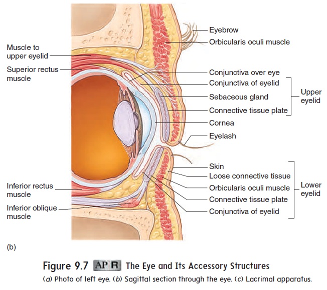
Accessory Structures Of The Eye

Accessory Structures Of The Eye

Senses Quiz Flashcards Quizlet
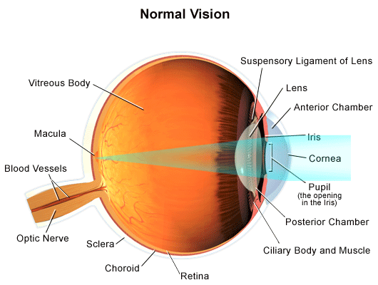
Normal Vision

Pin On Health Info 2

Compact Bone

Special Senses Hearing Audition And Balance A P 1 2 Senses Teaching Anatomy

Eye Diagram By Firkin Human Eye Diagram Diagram Of The Eye Eye Structure

Major Ocular Structures Laramy K Independent Optical Lab Freeform Lenses And Ar Coatings

Labeled Eye Diagram Human Eye Diagram Eye Anatomy Diagram Of The Eye

Help Students Understand How To Draw Lewis Structures Understand Molecular Geometry And The Polar Nature Of Ion Molecular Shapes Molecular Geometry Molecular

Eye Structures Front And Side Views

Anatomy Of The Human Eye


Comments
Post a Comment